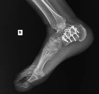
A middle-aged male construction worker presented with persistent right ankle pain and instability following a work-related injury. The patient sustained a right calcaneus fracture after jumping from a collapsing trailer while unloading materials. His injury occurred while working as an ironworker, and he required open reduction and internal fixation (ORIF) of the right heel at a tertiary medical center.
Despite initial healing, he continued to experience progressive ankle pain, stiffness, and functional impairment. Conservative management—including bracing, anti-inflammatory medications, and physical therapy—was attempted over several months but yielded limited relief. He reported increasing difficulty walking, swelling, and pain with weight-bearing activities, which interfered with his ability to work.
Diagnostic Workup and Preoperative Evaluation
After continued symptoms, imaging revealed:
Post-traumatic subtalar osteoarthritis following calcaneus ORIF
Tendonitis affecting the peroneal tendons
Persistent subtalar stiffness with pain exacerbated by movement
Given the failure of non-operative management, the patient was counseled on surgical intervention. Risks, benefits, and alternatives were discussed at length. Due to his ongoing functional limitations and worsening symptoms, he elected to undergo right subtalar arthrodesis with hardware removal and bone grafting.
Surgical Procedure
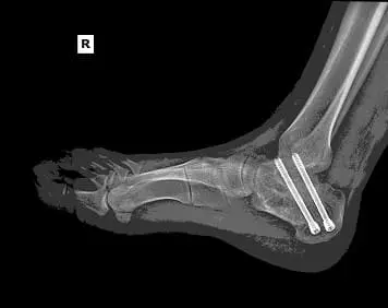
The patient underwent right subtalar arthrodesis with hardware removal at a local medical center. Intraoperatively, significant post-traumatic changes, joint degeneration, and residual deformity were observed. A structural bone graft was placed, and internal fixation was used to achieve stability. The procedure was successfully completed with minimal blood loss.
Postoperative Course and Rehabilitation
First Follow-Up
At his initial post-op visit, the patient was non-weight bearing (NWB) in a short-leg cast. He reported well-controlled pain but noted mild swelling. His incisions were clean and dry, with no signs of infection.
Pain management: Tylenol, Celecoxib, and Gabapentin were prescribed.
Follow-up X-rays: Confirmed hardware in situ with robust fusion progression.
Transition to Weight-Bearing
At subsequent visits, the patient progressed to a walking boot with partial weight-bearing (WBAT). He reported occasional swelling with prolonged activity but no severe pain. A structured physical therapy regimen was initiated, focusing on:
Range of motion exercises to improve mobility
Progressive weight-bearing drills
Proprioceptive training to restore balance and gait stability
By week 6 post-op, he was transitioned out of the boot and continued progressive loading of the ankle joint.
Final Stages of Recovery and Long-Term Outlook
At his three-month follow-up, the patient demonstrated significant functional improvement, with no residual pain in the subtalar joint. However, he developed shooting dorsal foot pain, likely due to lumbar radiculopathy or peripheral nerve irritation.
An electromyography (EMG) study was ordered to assess potential nerve involvement.
Conservative pain management, including Diclofenac gel and Gabapentin, was initiated.
By the final follow-up, he was fully weight-bearing, ambulating independently, and had resumed daily activities. Although mild residual stiffness and swelling persisted, he reported no limitations in basic mobility.
Conclusion
This case highlights the successful surgical management of post-traumatic subtalar arthritis following a calcaneus fracture. The combination of subtalar arthrodesis, hardware removal, and targeted rehabilitation allowed the patient to regain functional independence.
At the final evaluation, he:
Achieved stable joint fusion with no complications
Resumed full weight-bearing with minimal discomfort
Returned to daily activities without significant limitations
His outcome underscores the importance of timely surgical intervention and structured rehabilitation in restoring function after complex post-traumatic ankle injuries.
