Knee pain can make everyday activities difficult, especially when swelling or tenderness is present. In these situations, it is important to meet with a specialist. The knee is a complex joint made of bones, ligaments, tendons, muscles, cartilage, and fluid-filled structures that work together to support motion and weight bearing. Understanding how these parts fit and move helps explain why knee problems are so common and how they are treated.
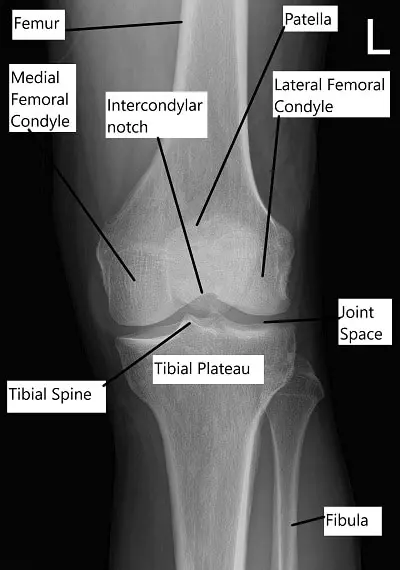
X-ray showing knee anatomy.
Functional Anatomy – Bones, Ligaments, Tendons, and Soft Tissues
The knee is formed by four bones:
Femur (thighbone), tibia (shinbone), patella (kneecap), and fibula (a thin bone alongside the tibia).
The upper ends of the tibia and femur create the main knee joint, while the patella sits in front and glides as the knee bends. The joint is lined by synovium, which makes fluid that nourishes the joint.
Ligaments hold the bones together:
- ACL limits forward movement of the tibia.
- PCL limits backward movement.
- MCL stabilizes the inner knee.
- LCL stabilizes the outer knee.
Tendons connect muscles to the knee. The quadriceps tendon links the front thigh muscles to the kneecap, and the patellar tendon connects the kneecap to the shinbone. The hamstrings, located behind the thigh, help bend the knee.
The menisci are two C-shaped cartilage pads, one on the inner knee and one on the outer knee. They act as shock absorbers.
Medial Meniscus: Located on the inner side of the knee.
Lateral Meniscus: Located on the outer side of the knee.
The menisci act as shock absorbers, reducing the impact on the knee joint during activities like walking and running.
Small fluid-filled sacs called bursae reduce friction. Important ones include the prepatellar, infrapatellar, and pes anserine bursae.
Prepatellar Bursa: Located in front of the patella.
Infrapatellar Bursa: Found below the patella.
Pes Anserine Bursa: Located on the inner side of the knee.
The knee joint is enclosed in a capsule that provides support and stability. The inner lining of the capsule, called the synovial membrane, produces synovial fluid, which lubricates the joint and reduces friction.
Biomechanics or Physiology – How Forces Move Through the Knee
The knee functions like a hinge, allowing bending and straightening. It also permits small amounts of rotation, especially when bent. The menisci and cartilage help spread weight evenly, protecting the joint surfaces. Every step, squat, or jump places load on the knee, making proper alignment and smooth gliding essential for pain-free movement.
Common Variants and Anomalies
Natural differences in knee shape, cartilage thickness, and alignment can affect how forces travel through the joint. Some people have bow-legged or knock-kneed alignment, which may place uneven strain on the inner or outer joint surfaces. The shape of the menisci and the depth of the tibial plateau also vary from person to person.
Clinical Relevance – How Dysfunction Leads to Pain or Injury
This joint structure allows powerful movement but also makes the knee vulnerable. Damage to cartilage can lead to arthritis. Ligament injuries can cause instability. Meniscal tears can result from twisting or sudden motion and may cause locking or catching. Irritated tendons can lead to pain during activity. When one structure is injured, it often affects the function of others.
Imaging Overview
X-rays show bone structure, alignment, and arthritis.
MRI shows cartilage, ligaments, tendons, menisci, and other soft tissues.
These images help identify tears, inflammation, instability, or joint wear.
Associated Conditions
Common knee-related conditions include:
- ACL tears from sudden stops or direction changes.
- Meniscal tears from twisting or impact.
- Patellar tendinitis from repetitive stress.
- Bursitis from friction or direct pressure.
- Arthritis from cartilage wear.
These problems can appear in athletes, older adults, or anyone with overuse or injury.
Surgical or Diagnostic Applications
Understanding knee anatomy helps surgeons plan procedures such as:
- ACL reconstruction
- Meniscus repair
- Patellar tendon repair
- Cartilage restoration procedures
- Total knee replacement in severe arthritis
Diagnostic steps may include X-rays, MRI, or joint fluid analysis depending on symptoms.
Prevention and Maintenance
Knee health can be supported through:
- Strengthening quadriceps, hamstrings, and calf muscles
- Stretching to maintain flexibility
- Wearing supportive shoes
- Avoiding overuse by pacing activity
- Using proper form during sports or exercise
These approaches help protect the joint surfaces and prevent strain.
Research Spotlight
A recent study evaluated how restoring the medial knee anatomy during total knee arthroplasty affects stability and muscle function, showing that a technique designed to better reproduce the knee’s natural joint line did not reduce mid-flexion laxity but did improve quadriceps peak force compared with the standard approach.
This finding reinforces the importance of understanding normal knee structure—bones, ligaments, menisci, cartilage, and supporting muscles—because even small variations in joint alignment can influence how well the knee functions after surgery.
While the clinical presentation of knee pain and the need for accurate diagnosis remain rooted in core anatomy such as the femur, tibia, patella, and major stabilizing ligaments, the study highlights that surgical techniques aiming to recreate native knee mechanics may enhance muscle performance without necessarily changing overall stability.
These insights support careful attention to alignment and soft-tissue balance during treatment for knee pain, whether caused by arthritis, ligament injury, or postoperative changes. (“Study of medial joint line restoration in total knee arthroplasty – See PubMed.”)
Summary and Key Takeaways
The knee is a complex joint designed for weight bearing and movement. It includes bones, ligaments, muscles, cartilage, and fluid-filled structures that work together smoothly when healthy. Because of its importance and constant use, it is prone to injury. Understanding knee anatomy helps explain symptoms and guides treatment, from simple strengthening to advanced surgical care.
Do you have more questions?
What are the main functions of the knee joint?
The knee joint allows for movement (flexion and extension), supports body weight, and provides stability during activities like walking, running, and jumping.
How does the ACL prevent knee injuries?
The ACL prevents the tibia from sliding forward and provides rotational stability, which is crucial during activities involving sudden stops or changes in direction.
What role do the menisci play in the knee joint?
The menisci act as shock absorbers, distribute weight evenly across the knee, and provide stability by improving the fit between the femur and tibia.
How do the quadriceps muscles contribute to knee function?
The quadriceps muscles straighten the knee (extension) and stabilize the patella, enhancing the knee’s ability to bear weight and perform activities.
What is the significance of the patellar tendon?
The patellar tendon connects the patella to the tibia, transmitting the force from the quadriceps muscles to straighten the knee.
How can one prevent common knee injuries?
Prevent injuries by strengthening muscles around the knee, maintaining flexibility, wearing proper footwear, and avoiding excessive stress on the knee joint.
What is the role of the synovial fluid in the knee?
Synovial fluid lubricates the knee joint, reducing friction and allowing smooth movement between the joint surfaces.
How does the PCL differ from the ACL?
The PCL prevents the tibia from sliding backward and provides stability in the posterior direction, while the ACL prevents forward sliding and rotational instability.
What causes patellar tendinitis, and how is it treated?
Patellar tendinitis, or “jumper’s knee,” is caused by overuse and repetitive stress. Treatment includes rest, ice, physical therapy, and sometimes anti-inflammatory medications.
Why are bursae important in the knee joint?
Bursae reduce friction and cushion the knee, preventing irritation and inflammation of the surrounding tissues during movement.
What types of exercises are beneficial for knee health?
Strengthening exercises for the quadriceps, hamstrings, and calf muscles, as well as flexibility and balance exercises, are beneficial for knee health.
How does cartilage contribute to knee function?
Cartilage covers the ends of bones in the knee, providing a smooth, lubricated surface for joint movement and acting as a cushion to absorb impact.
What is the function of the MCL and LCL in knee stability?
The MCL provides stability against inward forces, while the LCL provides stability against outward forces, both crucial for maintaining knee alignment.
How do rotational movements affect the knee joint?
Rotational movements can stress the knee ligaments, particularly the ACL, increasing the risk of injury if the knee is not adequately stabilized.
What are common symptoms of a meniscal tear?
Symptoms include pain, swelling, stiffness, and a clicking or locking sensation in the knee.
How is an ACL tear diagnosed and treated?
An ACL tear is diagnosed through physical examination and imaging tests like MRI. Treatment may include rest, physical therapy, and surgery for severe cases.
What factors contribute to knee osteoarthritis?
Contributing factors include aging, joint injury, repetitive stress, obesity, and genetics.
How does knee anatomy differ between children and adults?
Children’s knee anatomy is still developing, with growth plates present in the bones, making them more susceptible to certain types of injuries compared to adults.
What is the recovery process for knee ligament injuries?
Recovery involves rest, physical therapy, and gradual return to activity. Severe injuries may require surgical repair and extensive rehabilitation.
How can proper footwear protect the knees?
Proper footwear provides support, cushioning, and stability, reducing the impact on the knees and preventing injuries.
What are the signs of a PCL injury?
Signs include pain, swelling, and instability in the knee, especially when bearing weight or walking downhill.
Why is knee flexibility important?
Flexibility allows for a full range of motion, reducing the risk of stiffness and injury, and ensuring proper function of the knee joint.
How do knee braces help in injury prevention and recovery?
Knee braces provide support, reduce stress on the knee, and limit movement to prevent further injury and aid in recovery.
What are the long-term effects of untreated knee injuries?
Untreated knee injuries can lead to chronic pain, instability, reduced mobility, and an increased risk of developing osteoarthritis.
How does weight affect knee health?
Excess weight puts additional stress on the knee joint, increasing the risk of injury and accelerating the wear and tear on cartilage and other structures.